Programs


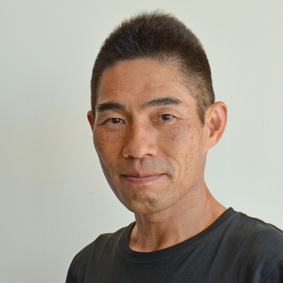
Professor Kenji Doya

Okinawa Institute of Science and Technology (OIST) Graduate University, Japan

TITLE: Brain/MINDS 2.0 and the Digital Brain Project

Abstract: Following the conclusion of the Brain/MINDS project (2014-2024), a new six-year program Multidisciplinary Frontier Brain and Neuroscience Discoveries (Brain/MINDS 2.0) has started. A remarkable feature of this program is that the Digital Brain plays a central role in integrating structural and dynamic brain data from multiple species for understanding brain functions and tackling neuropsychiatric disorders. This talk will present what is the Digital Brain of Brain/MINDS 2.0, how we can build that, and how we can use that. The primary aims of the Digital Brain Project are to develop open-source software tools for data-driven model building by integrating anatomical, genetic, physiological, and behavioral data from mice, marmosets, macaques and humans and to provide cloud-based platform for cross-species data search, data-driven model building, and simulation analyses. By utilizing those tools and platforms, we aim to build models that realize brain functions like reinforcement learning and Bayesian inference and reproduce neurodegenerative disorders like Parkinson’s disease and psychiatric disorders like schizophrenia to help early diagnosis and exploration of therapeutic and preventive strategies. This ambitious project requires fresh talents from math, computation, AI and brain sciences, as well as broad international collaborations. Through this conference, we hope to extend our network with researchers and research projects with overlapping interests and technologies.

Biography: Kenji Doya is a Professor of Neural Computation Unit, Okinawa Institute of Science and Technology (OIST) Graduate University. He studies reinforcement learning and probabilistic inference, and how they are realized in the brain. He took his PhD in 1991 at the University of Tokyo, worked as a postdoc at U. C. San Diego and the Salk Institute, and joined Advanced Telecommunications Research International (ATR) in 1994. In 2004, he was appointed as a Principal Investigator of the OIST Initial Research Project and as OIST established itself as a Graduate University in 2011, he became a Professor and served as the Vice Provost for Research till 2014. He served as a Co-Editor in Chief of Neural Networks from 2008 to 2021 and the Chairperson of Neuro2022 in Okinawa, and currently serves as the President of Japanese Neural Network Society (JNNS). He received INNS Donald O. Hebb Award in 2018, JNNS Academic Award and APNNS Outstanding Achievement Award in 2019, and the age-group 2nd place at Ironman Malaysia in 2022.

Professor Hanchuan Peng

SEU-ALLEN Joint Center, Southeast University, China

TITLE: Toward Building a Whole Brain Connectome at Single Neuron Resolution

Abstract: In this talk I will discuss our work of a large-scale study of whole-brain morphometry, analyzing 3.7 peta-voxels of mouse brain images at the single-cell resolution, producing one of the largest multi-morphometry databases of mammalian brains to date. We annotated 3D locations of cell bodies of 182,497 neurons, modeled 15,441 dendritic microenvironments, characterized the full morphology of 1,876 neurons along with their axonal motifs, and detected 2.63 million axonal varicosities that indicate potential synaptic sites. Our analysis covers six levels of information related to neuronal populations, dendritic microenvironments, single-cell full morphology, sub-neuronal dendritic and axonal arborization, axonal varicosities , and sub-neuronal structural motifs, along with a quantification of the diversity and stereotypy of patterns at each level. Overall, our study provides an integrative description of key anatomical structures of neurons and their types, covering a wide range of scales and features, and contributes a large-scale resource to understanding neuronal diversity in the mammalian brain. With this dataset, we start to formulate a possible whole brain scale connectome at the single neuron resolution for mouse brains.

Biography: Hanchuan Peng (Fellow, IEEE, AIMBE, Founding Director – Institute for Brain and Intelligence; Founding Director – SEU-ALLEN INSTITUTE Joint Center) develops technologies to generate, manage, visualize, analyze, and understand massive-scale structure and function data related to brains and other biomedical applications. Peng was the Director – Advanced Computing, Allen Institute for Brain Science, and also an Affiliate Professor with University of Washington, University of Georgia, among others). Peng was also a PI with Janelia, HHMI. Peng’s original work include the widely cited mRMR feature selection algorithm, APP1/APP2 neuron reconstruction algorithms, Virtual Fingers, Vaa3D, TeraFly, TeraVR, etc. His workwas cited about 30,000 times, in a number of fields. Peng founded Bioimage Informatics conferences in 2005, and iconized Bioimage Informatics as a new field in major bioinformatics journals including Bioinformatics, BMC Bioinformatics, Nature Methods, Nature Biotechnology, etc. He was the co-Editor-in-Chief of Brain Informatics (2016-2020) and a Section Editor of BMC Bioinformatics (2011-2018), and Bioinformatics (2021-2024).


Professor Allison Sekuler

Baycrest Health Sciences, McMaster University & University of Toronto, Canada

TITLE: Predictive Neuroscience for Precision Aging: Dementia Prevention, Detection, Treatment, and Care

Abstract: The world is aging faster now than ever before, and as we age the risk of dementia is growing. Brain changes linked to Alzheimer’s Disease (AD) and related dementias begin years before the onset of clinical symptoms. However, we lack sufficient tools to accurately identify individuals during preclinical stages of dementia, which limits our ability to implement interventions that could prevent or slow disease progression. Although memory loss is one of the most common symptoms of dementia, visual perceptual and attention can also be impacted at the early stages of disease, but can be difficult to assess throughout disease progression. I will describe some of the ways in aging affects visual perception (including behavioural assessments and electrophysiological markers of face processing and contour integration), how those changes differ in healthy aging and neurodegeneration, and how tools from vision science can probe function in individuals living with dementia and beyond. The results of the work I will discuss are important for developing rapid, non-verbal assessments of visual function that could be used as early screening tools for dementia and assessment throughout disease progression. I also will share other examples of Baycrest’s approach to predictive neuroscience for precision aging, including a range of collaborative opportunities spanning dementia prevention, detection, treatment, and care.

Biography: Dr. Allison Sekuler (FSEP, FPS, FAPS) is the Sandra A. Rotman Chair in Cognitive Neuroscience and Vice-President Research at Baycrest Health Sciences. A graduate of Pomona College (BA, Mathematics and Psychology) and the University of California, Berkeley (PhD, Psychology), Dr. Sekuler holds faculty positions in the Department of Psychology, Neuroscience & Behaviour at McMaster University and the Department of Psychology at the University of Toronto. Her research uses behavioural and neuroimaging approaches to understand how the brain processes visual information, with specific interests in face perception, motion processing, perceptual learning, neural plasticity, aging, and neurotechnology. Her research was the first to show conclusively that older brains “rewire” themselves to compensate for functional changes. Her clinical and translational research aims to develop methods to prevent, detect, and treat age-related sensory and cognitive decline. She has scientific and industry collaborations across North America, the EU, and Asia, and her work has been published in leading international journals, including Nature, Current Biology, and the Journal of Neuroscience.
She Chairs the Natural Science and Engineering Research Council’s Public Impact Value Proposition committee; serves on the Board of Governors for Hamilton Health Sciences and BrainsCAN and the scientific advisory board for VISTA; and is a founding steering committee member of the Canadian Brain Research Strategy. She also is a longstanding and passionate supporter of research communication and public outreach, serving, for example, as the only scientist on founding committee of the the Science Media Centre of Canada; and a sought-after speaker, podcaster, and commentator in national and international media. Co-founder of FoVea (Females of Vision et al.), an international organization to advance women in vision science, and co-Executive Champion of the Ontario Hospital Association’s Research and Innovation Anti-Racism Taskforce, Dr. Sekuler is a highly respected advocate for women and underrepresented groups in science, engineering, and technology. Dr. Sekuler has won numerous national and international awards for research, teaching, and leadership -- including serving as the country’s first Canada Research Chair in Cognitive Neuroscience and recently being named one of WXN's Top 100 Most Powerful Women in Canada (2019). In her spare time, she is proving that you’re never too old to learn: she picked up her first drumsticks a few years ago, joined a band, and recently earned her Drum Professional Certificate from the Berklee College of Music.


Professor Ramesh Srinivasan

University of California, Irvine, USA

TITLE: Graphical Modeling of Brain Networks

Abstract: Much of our understanding of human brain function is developed from the analysis of statistical relationships between brain signals and behavior. Graphical models of brain signals are generative models that potentially provide causal insight into brain signals and their relationship to behavior and disease. I will discuss different studies in graphical modeling that we have used to (1) model structure-function relationships, (2) model the relationship between brain injury and function, (3) develop new approaches to hyperscanning based on symbolic dynamics, (4) model joint latent space to link cognitive parameters to both neural signals and behavioral measures. To study structure-function relationships we incorporate anatomical knowledge of brain networks to build a graphical model of brain signals and demonstrate in fMRI data that we can predict the effects of disconnection due to injury in stroke (Wodeyar et al., 2021). These graphical models capture the dynamic effects of injury in a manner not apparent in anatomy or in the raw signals. Measures of network properties in structurally informed graphical models of EEG reflect how efficient signal routing is essential to maintain motor functional status after stroke(Zhou et al., 2024). Graphical modeling also provides an entirely new approach to hyperscanning in coordination and other forms of social cognitive neuroscience. We modeled the joint state of two individuals performing coordinated motor tasks with simultaneous EEG recordings, as a transition network in a symbol space defined by the graphical models, i.e., a graph of graphs. The symbolic dynamics over this graphical model capture the different coordination modes in a manner not possible by statistical analysis of correlations between brain signals. Graphical modeling can also be useful for formulating the link between brain activity and latent cognitive processes. Behavioral measures, such as accuracy and speed of motor responses, reflect latent cognitive processes underlying decision making. We have developed a novel approach that allows a theoretical account of the cognitive process of decision-making, and artificial neural networks to estimate a joint latent space to link cognitive parameters to both neural signals and behavioral measures (Vo et al., 2024). This joint latent space model is a valuable new framework for computational cognitive neuroscience, allowing for new forms of inference and hypothesis generation. The power of graphical modeling can allow for a more comprehensive understanding of the triplet relationship between behavior, brain activity, and cognitive processes.

Biography: Ramesh Srinivasan is a Professor of Cognitive Sciences and Biomedical Engineering at the University of California, Irvine. The primary focus of Srinivasan’s research is on developing signal processing and computational modeling to relate brain networks to cognitive functions. This foundational signal processing research has broad impact in clinical and cognitive neuroscience research. Dr Srinivasan’s PhD training in Biomedical Engineering at Tulane University was on the development of theoretical models of electroencephalography (EEG) largely contributed to the book (with Paul Nunez) Electric Fields of the Brain: The Neurophysics of EEG, 2nd ed., Oxford UP . He developed cognitive science and neuroscience expertise through postdoctoral training at the University of Oregon and at the Neurosciences Institute in San Diego. He joined the faculty in Cognitive Sciences and Biomedical Engineering at the University of California, Irvine in 2000 and was Chair of the Cognitive Sciences department from 2012 to 2022. He was appointed a Senior Fellow of the US Army Research Lab in 2022. He has published more than 100 papers in EEG/MEG signal processing, theoretical computational neuroscience, cognitive neuroscience applications in perception, attention, and decision making, and clinical research especially focused on motor functions and stroke. His recent work is focused on graphical models of EEG, MEG, and fMRI signals that incorporate structural and functional modeling to develop probabilistic generative models of brain networks linked to behavior and clinical status.


Professor Lucina Q. Uddin

University of California Los Angeles, USA

TITLE: Neuroinformatics and Cognitive Ontologies

Abstract: Decades of cognitive neuroimaging work has identified distinct patterns of brain activation that occur during performance of different tasks, as well as revealed patterns of task-general activation and deactivation. These data can in principle be used to provide the basis for constructing biologically informed, data-driven taxonomies of psychological processes. This talk will highlight some of the progress and challenges associated with the construction of cognitive ontologies based on functional neuroimaging data.

Biography: After receiving a Ph.D. in cognitive neuroscience from the Psychology Department at the University of California Los Angeles, Dr. Uddin completed a postdoctoral fellowship in the Child Study Center at New York University. For several years she worked as a faculty member in Psychiatry & Behavioral Science at Stanford University. She recently returned to UCLA where she currently directs the Brain Connectivity and Cognition Laboratory and the Center for Cognitive Neuroscience Analysis Core in the Semel Institute for Neuroscience and Human Behavior. Within a cognitive neuroscience framework, Dr. Uddin's research combines functional and structural neuroimaging to examine the organization of large-scale brain networks supporting the development of social cognition and executive function. Her current projects focus on understanding dynamic brain network interactions underlying cognitive inflexibility in neurodevelopmental conditions such as autism spectrum disorder. Dr. Uddin's work has been published in the Journal of Neuroscience, Cerebral Cortex, JAMA Psychiatry, Biological Psychiatry, PNAS, and Nature Reviews Neuroscience. She was awarded the Young Investigator award by the Organization for Human Brain Mapping in 2017.